Traumatic brain injury (TBI) management in Pune is available at Manipal Hospital, Baner, under Dr. Sarang Gotecha, MCh Neurosurgery. He manages all grades of TBI - from observation for mild concussion to emergency craniotomy for epidural or subdural haematomas - using ICP monitoring, neuronavigation and a dedicated neuro-ICU.
Lead with a local and national fact: Road traffic accidents are the leading cause of traumatic brain injury in India, accounting for over 60% of TBI admissions in urban trauma centres. Pune, with its high traffic density and large two-wheeler population, sees a disproportionately high volume of head injuries. The critical insight: the outcome of a TBI patient is determined largely by where they are taken in the first 60 minutes after injury. Being in proximity to Manipal Hospital, Baner, matters.
Every hour, someone in Pune suffers a significant head injury. Most are from road accidents - two-wheelers without helmets, vehicles colliding at speeds that the skull and brain are not built to withstand. Others come from falls - the elderly patient who falls at home, the construction worker who falls from scaffolding.

The management of traumatic brain injury (TBI) begins at the scene of the accident and continues through the emergency department, operating theatre, ICU and rehabilitation. Getting each phase right is what determines who walks out of the hospital and who does not.
Dr. Sarang Gotecha manages TBI cases at Manipal Hospital, Baner - a facility with the full infrastructure required for TBI care: CT scanner, neurosurgical operating theatre, neuro-ICU and a dedicated neurosurgical team available around the clock.
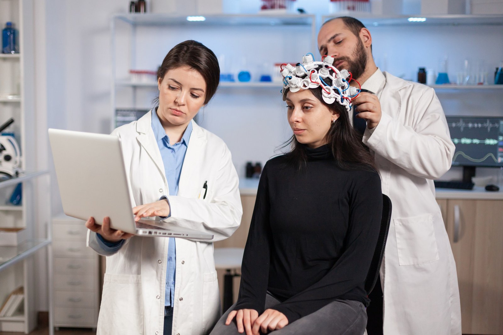
TBI occurs when external physical force causes damage to the brain. This force may cause the brain to strike the inside of the skull (contusion), disrupt blood vessels (haematoma), damage axons (diffuse axonal injury), or cause the brain to swell (cerebral oedema).
The skull, designed to protect the brain from normal daily impacts, provides no protection against the forces generated in road accidents or high-impact falls. The rigid closed box of the skull means that any swelling or bleeding inside it creates pressure - and pressure on the brain causes damage.
Includes concussion. Brief loss of consciousness (< 30 minutes) or none, confusion, amnesia for the event. Most patients recover fully with observation and rest. CT is performed to rule out intracranial bleeding.
Moderate TBI (GCS 9–12):Longer loss of consciousness, confusion lasting hours, often with CT abnormalities (contusions, small haematomas). Hospital admission for observation and serial neurological assessment is standard.
Severe TBI (GCS 3–8):Life-threatening injury. The patient cannot follow commands or speak coherently. Emergency CT scanning, neurosurgical consultation and often emergency surgery or ICP monitoring are required. Mortality in severe TBI without surgical treatment of treatable lesions exceeds 50%.
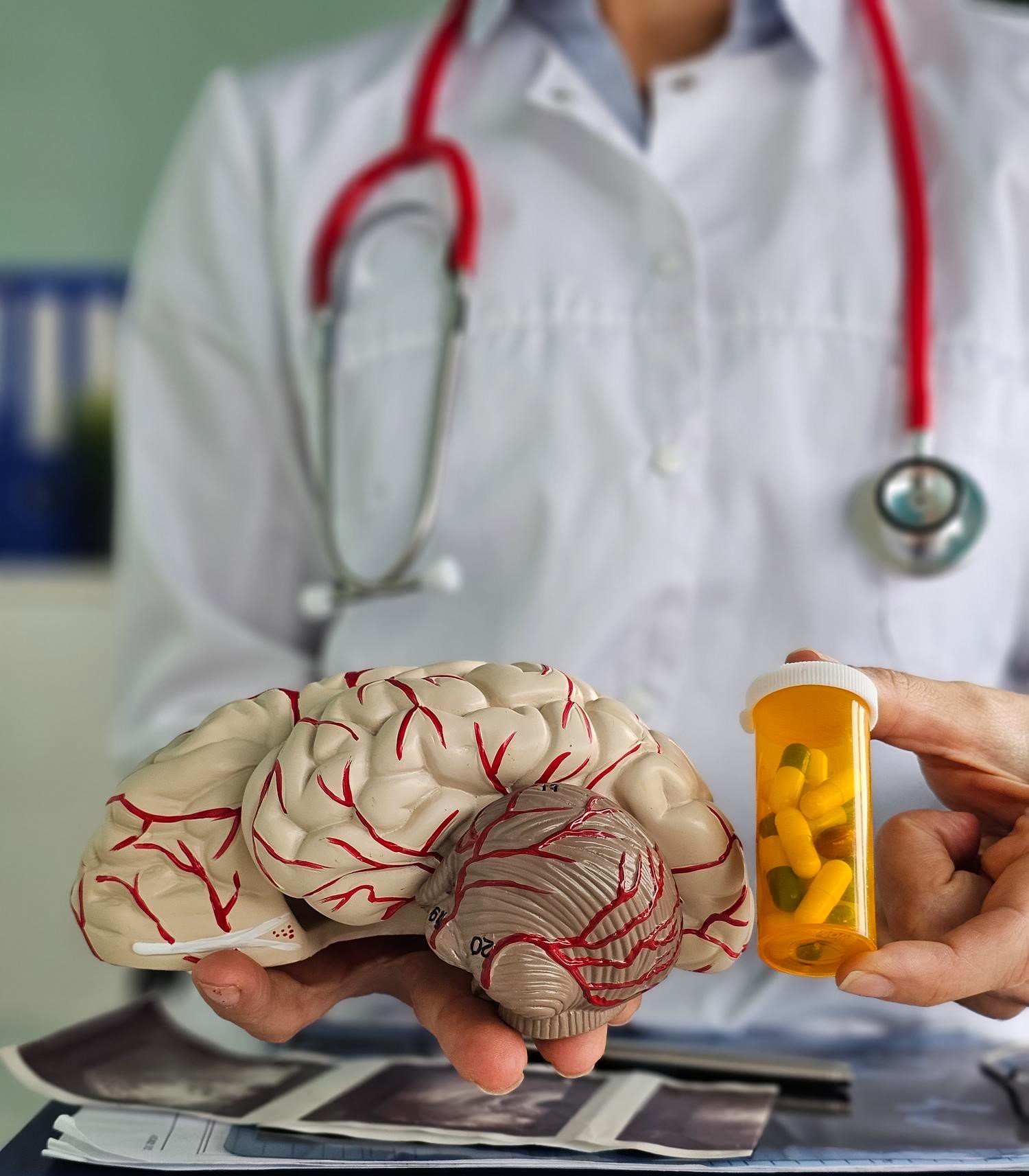
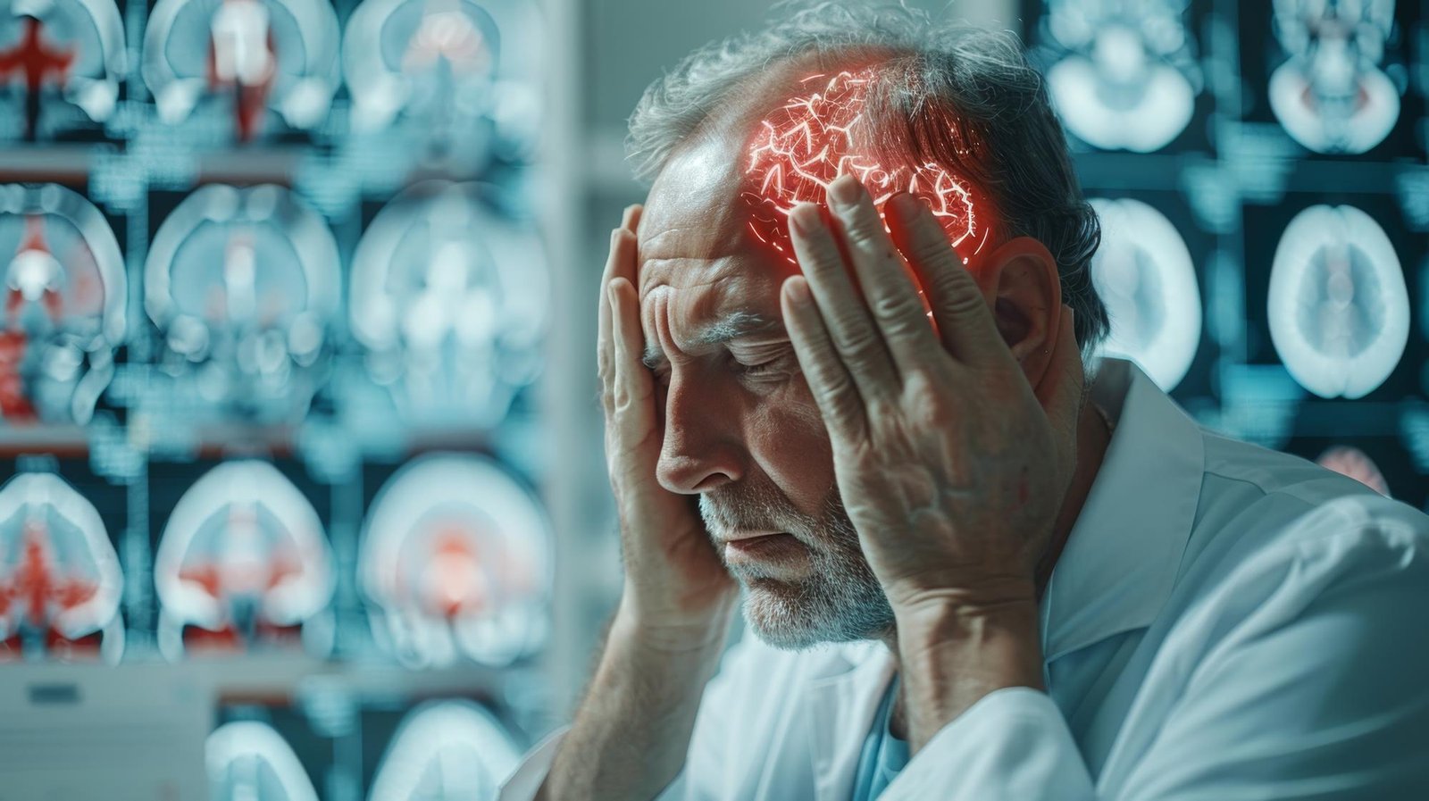
An epidural haematoma is a collection of blood between the skull and the outer brain covering (dura). It typically results from a skull fracture tearing the middle meningeal artery. The classic presentation - a lucid interval after the initial impact, followed by rapid neurological deterioration - is a neurosurgical emergency. Emergency craniotomy and haematoma evacuation is life-saving and, if done before brain herniation, carries an excellent prognosis.
A subdural haematoma is a collection of blood between the dura and the brain surface, caused by tearing of bridging veins. Acute SDH (< 72 hours post-injury) with significant brain compression requires emergency surgical evacuation - craniotomy or burr holes depending on the blood's consistency and the patient's clinical status. Chronic SDH (weeks after injury, often in elderly patients on blood thinners) can be drained through burr holes under local or general anaesthesia.
Traumatic intracerebral haemorrhage - bleeding within the brain substance - may be managed surgically or conservatively depending on the haematoma's size, location and the patient's neurological status. Haematomas causing significant midline shift or progressive neurological decline are evacuated surgically.
A fragment of skull driven into the brain (open or closed depressed fracture) requires surgical elevation and bone fragment removal when depression exceeds the thickness of the skull, there is dural laceration, or underlying brain injury is present. Infection risk is significantly higher with open fractures.
• Intracranial pressure (ICP) monitoring - a probe placed in the brain or ventricle continuously measures pressure
• Cerebral perfusion pressure (CPP) optimisation - ensuring the brain receives adequate blood flow despite elevated ICP
• Controlled ventilation, head positioning, osmotherapy (mannitol, hypertonic saline)
• Sedation and analgesia to reduce brain metabolic demand
ICP monitoring is available at Manipal Hospital, Baner and forms a core part of Dr. Gotecha's severe TBI management protocol.
Any patient who presents with significant head injury, loss of consciousness, amnesia, vomiting, or worsening headache after a head impact needs an emergency brain CT scan. This is non-negotiable. A normal neurological examination does not rule out a life-threatening haematoma.
Brain CT scan cost in Pune at major hospitals ranges from Rs. 2,500 to Rs. 6,000. At emergency presentation, this cost is covered by most health insurance policies. Do not delay imaging for cost concerns when a head injury is involved.
The CT scan identifies haematomas, fractures, contusions, brain swelling and midline shift - all of which determine the management pathway. Dr. Gotecha reviews CT findings personally and forms an immediate management plan.
TBI recovery is measured in months, not weeks. The brain's capacity for plasticity - rerouting function around injured areas - is real but requires time and appropriate rehabilitation.
Post-TBI rehabilitation includes physiotherapy (for motor deficits), speech and language therapy (for communication problems), occupational therapy (for daily activity retraining) and neuropsychological support (for cognitive and emotional sequelae).
Dr. Gotecha coordinates post-acute care at Manipal Hospital, Baner, with the rehabilitation medicine team, ensuring continuity from acute neurosurgical care to functional recovery. Follow-up consultations are available at Wakad and Thergaon clinics for patients in western Pune.
